Inflammation as a developmental signal
Maternal immune activation is associated with altered neurodevelopmental outcomes, but the mechanisms linking maternal inflammation to changes in the fetal brain are still being defined. Our work asks how specific inflammatory signals act directly on human neural stem cells and developing neural tissue.
We use human 3D experimental models to separate direct effects on the brain from the many systemic changes that accompany maternal inflammation in vivo. This allows us to test defined cytokines, identify responsive cell populations and follow downstream changes in tissue growth and organisation.
- Direct effects of IL-17A on human neural stem cells
- Changes in neurogenesis, neuronal maturation and cortical architecture
- NF-κB signalling as a downstream mechanism
- Interactions among neural, immune and vascular components of developing tissue
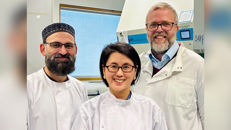
Our cerebroid work provides an experimental system for testing inflammatory effects directly in developing human brain tissue.
Recent finding
IL-17A disrupts cortical development in human cerebroids
Using ex vivo human fetal brain cerebroids, we found that IL-17A acts directly on neural stem cells and alters early cortical development. Exposure accelerated neuron production and maturation and produced changes in cortical tissue organisation, including premature folding.
The response was associated with sustained NF-κB signalling, and pharmacological inhibition of this pathway reversed many of the observed changes. These findings provide a mechanistic link between an inflammatory cytokine and altered developmental trajectories in human cortical tissue.
M. Z. K. Assir et al. Modeling maternal immune activation in 3D ex vivo human fetal brain cerebroids reveals IL-17A-driven disruption of cortical development. Nature Neuroscience, 2026. View publication
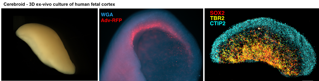
Cerebroids preserve native tissue complexity while permitting controlled cytokine exposure.
Why cellular context matters
Inflammatory responses are shaped by the cellular composition of the tissue. Our broader work examines whether microglia, endothelial cells and extracellular matrix remodelling influence the way developing neural tissue responds to IL-17A and related inflammatory signals.
This is one reason we use several complementary model systems rather than relying on a single organoid platform. Comparing models with different cellular complexity can help identify which components are required for a developmental phenotype to emerge.
Related research